A mole I'd had my entire life looked different in the mirror one morning. I'm high-risk for melanoma, and I knew what "different" could mean. The doctor agreed: very suspicious, biopsy it now. In the days that followed, waiting for my results, I felt ravaged with regret that so many months had gone by since I had done my last self skin-check. Melanoma is visible to the naked eye in its earliest stages. I felt angry that I had not been more diligent about my skin checks but also angry at the health care system. Luckily, it turned out not to be cancer, but I refuse to experience such regret again.
The screening problem
The current standard of care for skin cancer detection for high risk patients is woefully inadequate. A doctor inspects a patient’s skin once a year for a few minutes and reminds them that it’s their responsibility to check their skin regularly for any changes. This approach is poorly suited to melanoma, a cancer for which survival depends heavily on the stage at detection: five-year relative survival is greater than 99% for localized melanoma but falls to approximately 35% once the cancer has spread to distant organs.1National Cancer Institute. SEER Cancer Stat Facts: Melanoma of the Skin. Five-year relative survival based on SEER 21 data, 2016–2022. It also places the hardest part of the problem on the patient: early melanomas can appear as tiny, ambiguous changes in color, shape, or size, often hidden among dozens or hundreds of benign spots. Detecting them requires not merely looking at the skin, but remembering what every region looked like months earlier and recognizing which subtle changes matter.
The usual advice to “watch your moles” also understates the difficulty of the problem. Only about 30% of melanomas arise from an existing mole; roughly 70% appear as entirely new lesions on previously normal-looking skin.2Pampena R, Kyrgidis A, Lallas A, et al. A meta-analysis of nevus-associated melanoma: Prevalence and practical implications. Journal of the American Academy of Dermatology. 2017;77(5):938–945.e4. Tracking changes in every known mole is difficult. Recognizing that one tiny spot among hundreds of moles, freckles, and other benign features was not present six months earlier is even harder. A highly motivated patient can photograph their entire skin surface (I did this), but comparing those images over time is an enormous registration problem: differences in body position, camera angle, distance, lighting, and skin deformation make it difficult to align the same regions across scans. Finding a tiny new lesion therefore requires painstakingly matching and comparing hundreds of corresponding patches of skin.
The limits of total-body photography
We need a system that can create a high-resolution, repeatable map of the entire skin surface and automatically register each new scan to the last: total-body photography (TBP). Existing TBP systems such as Canfield’s VECTRA WB360 or Neko’s skin scan surround a standing patient with dozens of cameras, capture nearly the entire skin surface at once, and reconstruct a 3D model on which lesions can be mapped and tracked over time. This makes longitudinal surveillance far more systematic, particularly for people at high risk of melanoma. Yet these systems remain available primarily through a few specialized centers and are far from routine or broadly accessible. They are large and capital-intensive, and suspicious lesions must still be examined individually using higher-resolution dermoscopy. Most importantly, the evidence that adding 3D total-body photography improves melanoma detection over usual clinical surveillance remains mixed: a recent randomized trial found that the VECTRA WB360 did not increase the average number of melanomas detected.3Soyer HP, Jayasinghe D, Rodriguez-Acevedo AJ, et al. 3D Total-Body Photography in Patients at High Risk for Melanoma: A Randomized Clinical Trial. JAMA Dermatology. 2025;161(5):472–481., 4Lindsay D, Soyer HP, Janda M, et al. Cost-Effectiveness Analysis of 3D Total-Body Photography for People at High Risk of Melanoma. JAMA Dermatology. 2025;161(5):482–489.
Why, then, has total-body photography not transformed melanoma screening? The first limitation is image resolution. Fixed camera arrays are designed to capture the entire body quickly, so each lesion occupies only a small portion of the resulting image. The images may be sufficient to flag a spot as new or changing, but they generally do not preserve the fine structural detail available through close-range or dermoscopic imaging. This creates two problems. Clinically, every flagged lesion must still be located on the patient and inspected individually by a dermatologist, turning automated screening into a tedious follow-up workflow. Scientifically, the images collected during routine total-body scans may not contain enough detail to train models to recognize the earliest visible signs of malignancy. This limits both what current systems can conclude and the quality of the longitudinal datasets they generate.
A second, closely related limitation is the nature of the available training data. Most melanoma-detection datasets consist of close-up or dermoscopic images of lesions that clinicians had already selected as suspicious. Models trained on these datasets learn to answer, “Does this particular lesion look malignant?” They are not trained to answer the more useful screening question: “What has changed on this person’s skin since the last scan and are any of these changes likely early signs of melanoma?” Solving that problem will require large, standardized, high-resolution longitudinal datasets containing repeated images of the entire skin surface—including the enormous background of benign lesions and the rare melanomas as they first emerge.

Why this is a robotics problem
This is really a robotics problem. Capturing repeatable, high-resolution images requires moving a camera across the body’s contours while maintaining a consistent distance, viewing angle, focus, and lighting. A robot can systematically scan the entire skin surface with far greater precision and consistency than handheld photography. Its proprioception also provides an estimate of the camera pose for every image, creating a strong geometric prior for registering thousands of overlapping images and reconstructing the skin surface in 3D. Because the task is entirely non-contact, it also avoids many of the manipulation challenges that make other robotics problems difficult. Therefore, I decided that after finishing my PhD in robotics, I would build a robot to address this problem.
Building OpenDerm
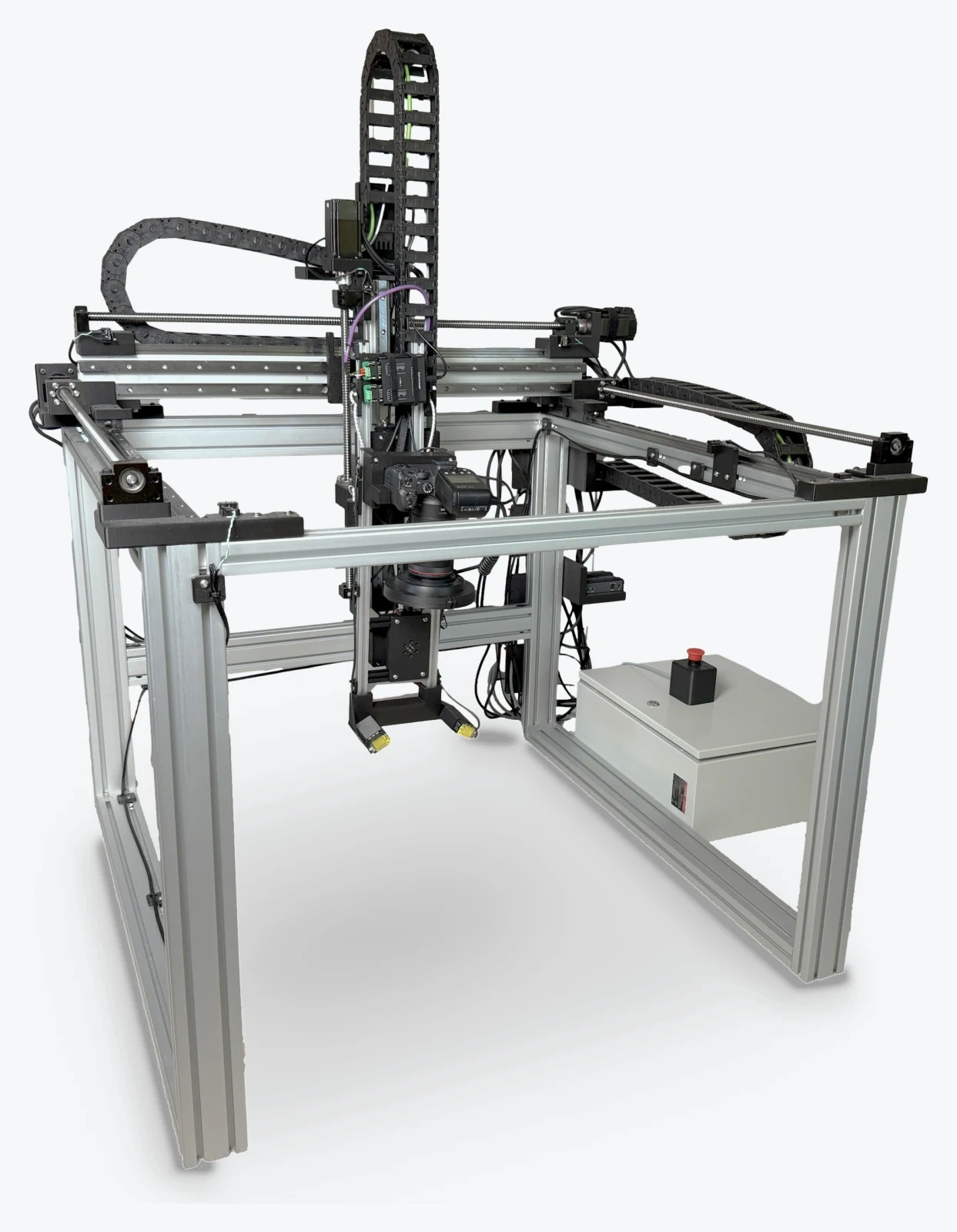
I built a 4-DOF robotic gantry called OpenDerm that moves a camera across the body with sub-millimeter positioning accuracy, capturing high-resolution, overlapping images of the skin. I built software that uses the robot’s estimated camera poses to register the images and reconstruct the scanned skin surface in 3D. Each new scan can then be registered against every previous scan, allowing the system to compare corresponding points across the skin surface and detect how individual lesions—and the surrounding skin—evolve over time.5I initially thought about using an off-the-shelf arm instead of building a custom robot. I went with the custom build approach because the task needs a large usable workspace more than dexterity. Most affordable arms cannot scan a full six-foot person while moving bulky imaging equipment around different angles. Larger arms could solve the workspace problem, but at a much higher price point. A custom gantry allowed me to optimize the robot’s workspace around the specific requirements of skin imaging rather than adapting the problem to an existing suboptimal and more expensive platform.
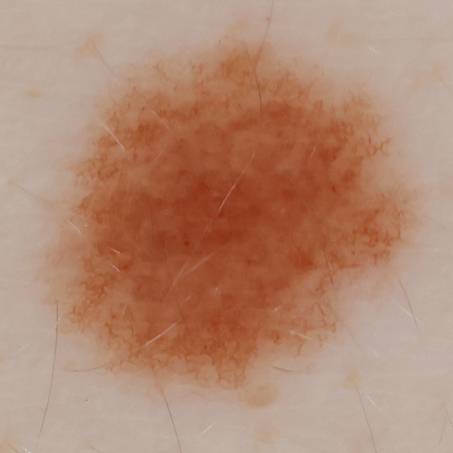
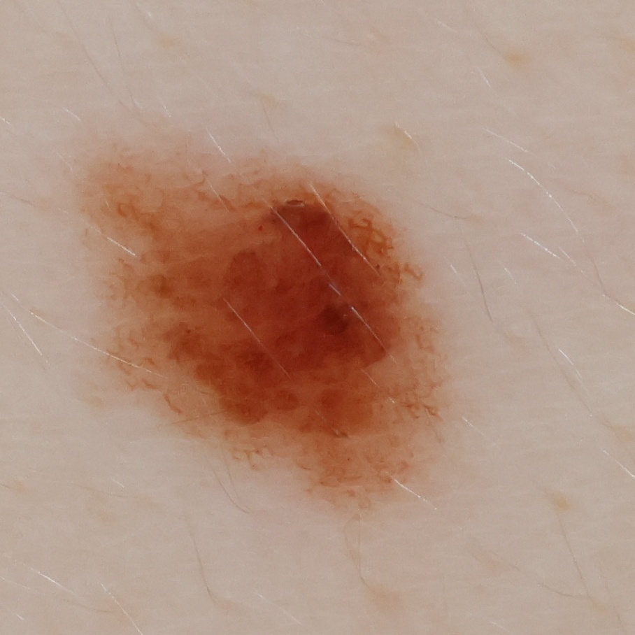
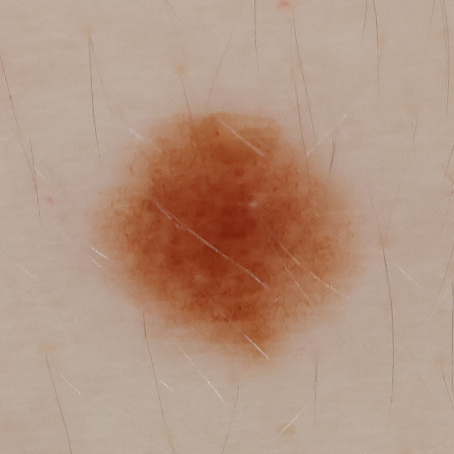
OpenDerm’s first prototype demonstrates that a robotic imaging system can capture the skin automatically at higher spatial resolution than wide-field total-body photography systems, and at a fraction of the cost. The system captures the skin at 78 pixels per mm and costs roughly $8,500 in parts.6There are obvious improvements for a v2 of this design. Most importantly: (1) the robot should scan the patient standing upright (rather than lying underneath it) for better user safety, and (2) it could be much faster with the gantry using belt-driven axes instead of ballscrews.
Skin imaging with general purpose robots
OpenDerm demonstrates a potential solution to several of the technical challenges of total-body photography, but it does not fully solve the access problem. Making the system open source lowers the barrier to reproducing it, but most people will not build a dedicated skin-imaging robot. OpenDerm is a valuable option for high-risk patients today, but the long-term answer for widespread adoption and data collection is not a dedicated imaging system.7In the interim, before widely-available personal robots, systems deployed by clinics and companies such as Neko could be an intermediary that make longitudinal scanning more widely available. Such deployments have the potential to generate valuable datasets, even if their wide-field cameras capture less detail than OpenDerm.
Ultimately, routine skin imaging belongs in every home. The same robot that cleans your house should also scan your skin.
A household robot could perform scans regularly, under consistent conditions, without requiring a special appointment or going to a dedicated imaging facility. That kind of low-friction, repeated measurement is the best way to bring longitudinal skin surveillance to everyone—catching skin cancer in its very earliest stages—and it is one of the reasons I’m so excited about the future of general-purpose robotics.
Footnotes
- National Cancer Institute. SEER Cancer Stat Facts: Melanoma of the Skin. Five-year relative survival based on SEER 21 data, 2016–2022. ↩
- Pampena R, Kyrgidis A, Lallas A, et al. A meta-analysis of nevus-associated melanoma: Prevalence and practical implications. Journal of the American Academy of Dermatology. 2017;77(5):938–945.e4. ↩
- Soyer HP, Jayasinghe D, Rodriguez-Acevedo AJ, et al. 3D Total-Body Photography in Patients at High Risk for Melanoma: A Randomized Clinical Trial. JAMA Dermatology. 2025;161(5):472–481. ↩
- Lindsay D, Soyer HP, Janda M, et al. Cost-Effectiveness Analysis of 3D Total-Body Photography for People at High Risk of Melanoma. JAMA Dermatology. 2025;161(5):482–489. ↩
- I initially thought about using an off-the-shelf arm instead of building a custom robot. I went with the custom build approach because the task needs a large usable workspace more than dexterity. Most affordable arms cannot scan a full six-foot person while moving bulky imaging equipment around different angles. Larger arms could solve the workspace problem, but at a much higher price point. A custom gantry allowed me to optimize the robot’s workspace around the specific requirements of skin imaging rather than adapting the problem to an existing suboptimal and more expensive platform. ↩
- There are obvious improvements for a v2 of this design. Most importantly: (1) the robot should scan the patient standing upright (rather than lying underneath it) for better user safety, and (2) it could be much faster with the gantry using belt-driven axes instead of ballscrews. ↩
- In the interim, before widely-available personal robots, systems deployed by clinics and companies such as Neko could be an intermediary that make longitudinal scanning more widely available. Such deployments have the potential to generate valuable datasets, even if their wide-field cameras capture less detail than OpenDerm. ↩